Have you ever wondered how scientists look inside cells or tissues with such incredible detail? One of the tools they use is a laser confocal microscope. This advanced type of microscope helps researchers capture high-quality images of tiny objects that are difficult to see with regular microscopes. The laser confocal microscope allows scientists to create clear, focused pictures and even 3D models of small structures like cells and tissues.
In this article, we’ll explore what a laser confocal microscope is, how it works, and why it’s so important for science.
What is a Laser Confocal Microscope?
A laser confocal microscope is a special type of microscope that uses lasers to scan a sample point by point, producing detailed images with high resolution.(1) Unlike traditional microscopes that shine light over the entire sample at once, the laser confocal microscope focuses light on just one specific point at a time. This focused light creates clearer images, especially for thick specimens.
The magic of the laser confocal microscope comes from its ability to scan different layers of a sample. By scanning at different depths, the microscope collects a series of images that can be combined to form a 3D picture. This makes it easier for scientists to study complex structures in detail.
How Does a Laser Confocal Microscope Work?
Let’s break down the process of how a laser confocal microscope works:
1. Laser Illumination:
The microscope uses a laser as the light source. The laser beam is focused to a single point on the sample, providing precise illumination. The laser scans across the sample point by point, which allows the microscope to create a detailed image.
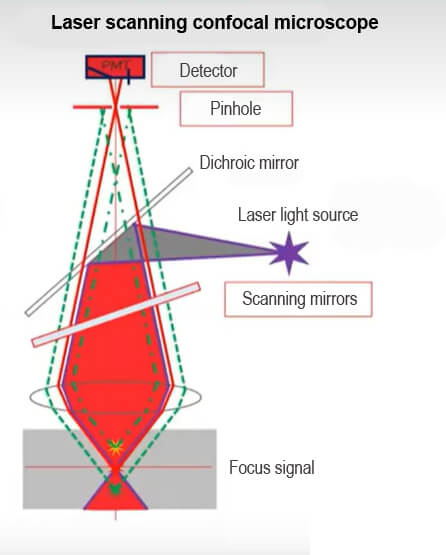
2. Galvo mirrors and Pinhole:
The laser beam passes through galvo mirrors and objective lens, which focuses the light onto the sample. After interacting with the sample, some of the light is reflected or emitted (if fluorescence is used). This reflected/emitted light is then collected by the same galvo mirrors and objective lens and directed through a small pinhole, which is the key to confocal imaging.
3. Pinhole Effect:
The pinhole is placed in front of the detector to block out-of-focus light from reaching it. Only light from the focal plane (the exact point where the laser is focused) passes through the pinhole. Light from above or below the focal plane is blocked, resulting in a sharp, high-contrast image with minimal background noise.
4. Image Acquisition and Scanning:
The laser beam is scanned across the sample in a raster pattern (horizontal and vertical movements), focusing on one point at a time. A detector, often a photomultiplier tube (PMT), measures the light intensity at each point and converts it into a digital signal.
5. 3D Imaging:
The laser confocal microscope can generate three-dimensional images by collecting data at different focal planes (optical sections) within the sample. By moving the focus of the laser up and down through the sample, a series of 2D images at different depths are captured. These images are then combined to create a 3D representation of the sample.
Why is the Laser Confocal Microscope Important?
The laser confocal microscope has several advantages that make it an essential tool in modern science:
- High Resolution: The microscope’s ability to focus on specific points and block out blurry light means it produces much sharper images than traditional microscopes. This is important when scientists need to study small details, like the structure of a cell or tissue.
- 3D Imaging: The laser confocal microscope can scan a sample at different depths, allowing researchers to build a 3D model of the object they are studying. This is incredibly useful for looking at complex structures, like how cells are arranged in a tissue or how different parts of a cell interact.
- Live Imaging: One of the amazing features of the laser confocal microscope is that it can be used to observe living cells without damaging them. This allows scientists to watch how cells move, divide, and interact in real time.
- Fluorescence Microscopy: Many laser confocal microscopes are used with fluorescent dyes that bind to specific parts of a cell. When exposed to the laser, these dyes light up, making it easy to see different components of the cell, such as DNA or proteins. This is called fluorescence microscopy and is often used in medical and biological research.
Key Components of a Laser Confocal Microscope
Here are some of the main parts that make a laser confocal microscope work:(2)
- Laser: The laser provides the light source. The focused beam of light scans the sample point by point, ensuring high-resolution images.
- Pinhole: This small opening blocks light from out-of-focus areas, allowing only in-focus light to pass through, which helps produce clearer images.
- Galvanometer-Controlled Mirrors: These mirrors direct the laser beam across the sample. They move quickly and precisely, allowing the microscope to scan the sample in all directions.
- Detector: The detector captures the reflected light and turns it into a digital image that can be analyzed by scientists.
Applications of the Laser Confocal Microscope
The laser confocal microscope is used in a wide variety of scientific fields, especially in biology and medicine. Here are a few key areas where it plays a vital role:
- Cell Biology: The laser confocal microscope is commonly used to study the inside of cells. Scientists can observe how different parts of the cell function and interact. This helps researchers understand diseases and how treatments work on a cellular level.
- Neuroscience: In brain research, the laser confocal microscope helps scientists study how neurons (nerve cells) are structured and how they connect with one another. This technology helps researchers understand brain function and disorders like Alzheimer’s disease.
- Cancer Research: Researchers use the laser confocal microscope to study how cancer cells grow and spread. It also helps in testing new treatments to see how cancer cells respond.
- Developmental Biology: Scientists can use the laser confocal microscope to study how organisms develop, watching how cells divide and form tissues in real-time.
Conclusion
The laser confocal microscope has revolutionized the way scientists study small structures like cells and tissues. Its ability to produce clear, high-resolution images and create 3D models makes it an invaluable tool in many areas of research. Whether it’s used to study cells, the brain, or cancer, the laser confocal microscope continues to help scientists make groundbreaking discoveries in the microscopic world.
REFERENCE
- https://en.wikipedia.org/wiki/Confocal_microscopy
- https://www.ncbi.nlm.nih.gov/pmc/articles/PMC6961134/